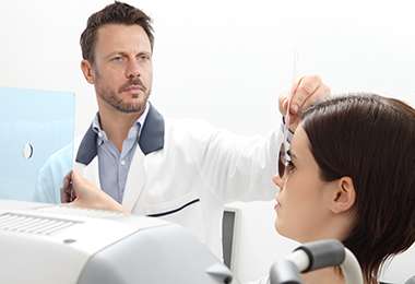
A Comprehensive Eye Exam is an important part in determining any possible eye conditions you may be experiencing. Many vision problems do not present any obvious symptoms, so waiting until you notice a change in your eyesight may delay much-needed treatment for a serious issue. The diagnostic tests that your ophthalmologist will perform will depend on your vision history, any current symptoms you are experiencing, and the doctor’s professional assessment. Below are some of the diagnostic tests that could be performed during your comprehensive eye exam.
Amsler Grid: An Amsler Grid test is used to diagnose macular problems when a patient complains of distorted vision, letters “jumping” off the page, or of an unexplained decrease in near vision. Patients describe the gridlines on a chart and any distortions noted are recorded by the technician.
Color Vision: A Color Vision test is used for screening whether a patient has acquired and/or hereditary color visual defects.
Corneal Topography: A Corneal Topography test maps the curvature of the cornea and is useful in diagnosing corneal diseases, such as an astigmatism or keratoconus (bulging of the cornea).
Binocularity (Cover Test): A Binocularity Test, or a Cover Test, is a test used to test the alignment of the eyes.
Dilation: The eyes are dilated using dilating (mydriatic) drops, which take about 15 minutes to take effect. Dilating the eye allows the ophthalmologist to see the back of the eye more clearly.
Dry Eye Test: A Dry Eye Test measures the amount of tears produced by using a special strip of paper that is placed under the lower lid for a certain amount of time.
Extended Ophthalmoscopy: An Extended Ophthalmoscopy test is used to see retinal detail. The ophthalmologist will use a special magnification.
External Exam: An External Exam consists of a CVF (Confrontation Visual Field), which gets a measurement of the peripheral vision, an EOM (Extraocular Motility), which measures the range of motion and the function of the extraocular muscles, and a Pupil Evaluation, which consists of evaluating the size, shape, and the pupil’s reaction to light.
Fundus Examination: A Fundus Examination occurs during a Slit Lamp Examination, where the ophthalmologist is able to examine the retina at the back of the eye. During the Fundus Examination, the ophthalmologist is able to determine the appearance of the optic nerve/disk, the macula, and the periphery and whether there are any abnormalities or diseases present.
Fundus Photography: Fundus Photography is photos of the appearance of the optic discs, the macula, and anomalies of the retina.
Glare Test – Brightness Acuity Testing (B.A.T.): A Glare Test determines the effects of glare on your visual acuity. This is often needed to document medical necessity for cataract surgery.
Gonioscopy: A Gonioscopy is primarily used as a screening for glaucoma. It is a mirrored contact lens evaluation that examines the angle structures in the front portion of the eye that allows for fluid outflow.
Keratometry: A Keratometry test measures the curvature of the cornea, which can be used to determine a fitting for an intraocular lens for cataract surgery, or to determine if there is a problem with the cornea.
Lensometry: A Lensometry measures a current glasses prescription.
Optical Coherence Tomography (OCT): An OCT test takes transpupillary images of the retina which assist in diagnosing retinal diseases.
Pachymetry: A Pachymetry is used to measure the thickness of the cornea, which is used in the diagnosis of glaucoma and corneal diseases.
Refraction: A Refraction determines the best prescription for glasses for any refractive errors of the eye.
Slit Lamp Examination: A Slit Lamp Examination is where a slit lamp, or biomicroscope, focuses a beam of light into the eye allowing the physician to examine the lids, eyelashes, conjunctive, cornea, iris, lens, vitreous, and fundus.
Stereopsis Test: A Stereopsis Test screens for normal/abnormal depth perception in the eyes.
Tonometry: A Tonometry measures the pressure of the eye.
Ultrasonography (Biometry): An Ultrasonography uses sound wave reflections or echoes to measure the length of the eye to detect abnormalities in the eye.
Visual Acuity: Visual Acuity is the number associated with the smallest objects a patient is able to see on specific eye charts from a specific distance. Normal visual acuity is considered 20/20.
Visual Field (Perimetry): A Visual Field is used to test for any defects in the central or peripheral vision.
and more!
Great staff. Knowledgeable and competent doctors. Two loved ones have personal experience with 2 of the doctors – office visits, cataract surgery. We’re very pleased.